Skin Lesions Requiring Surgical Treatment
What is a Skin Lesion?
Our skin has a more or less homogeneous appearance. Apart from physical injuries, there may be some formations that disrupt this smooth appearance. These lead to different properties, both visually and tactilely. For example, a raised appearance of skin attracts attention even if there is no color difference. Or a formation on the same level as the skin but in a different color draws attention. Even if there is no difference in color or appearance, if there is a difference in sensation, for example, if it hurts or itches, it still draws our attention. Each of these conditions is considered a skin lesion. Many times several findings occur together.
What causes skin lesions?
Skin lesions can be divided into two main parts
- Congenital lesions
- Lesions that happen later (acquired)
Congenital skin lesions
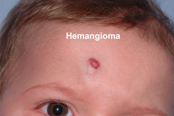
Almost all of these are benign formations. However, some rarely turn into bad habits (malignant) later in life. The most common ones can be under various groups such as vascular tumors (hemangioma), lymphatic system tumors (lymphangioma), nervous system tumors (neurofibroma), tumors of the cells that give color to the skin (congenital nevus). Most of these lesions cannot be treated with medication. If it is necessary, they are treated surgically.
Acquired (happened later) skin lesions
These lesions are seen in people of all ages and characterized by skin changes that occur at some point in life. As it has been mentioned earlier, these may be a new mass, discoloration, wounds of unknown origin, and painful and/or itchy conditions of the skin. If we group them:
- Sudden widespread skin lesions (skin manifestation of some diseases)
- Single lesion that grows slowly (usually a benign tumor)
- Single lesion that grows rapidly (a tumor that may be malignant, abscess)
- Lesions that suddenly appear and then disappear but reappear under certain conditions (allergy or some skin diseases)
Treatment of skin lesions
Multiple and sudden onset skin lesions suggest a systemic (involving the whole body) disease. Treatment should be directed towards the main disease. In single or a few skin lesions or tumors, surgical treatment is applied if medical (drug) treatment is not possible.
When should the skin lesion be surgically removed?
If a skin lesion remains in the same area for years without any change, it is usually benign. However, it may be requested to be removed for aesthetic reasons just because it creates visual discomfort and it is removed. In principle, every skin lesion removed, regardless of its appearance, must be sent for pathological examination. The reason will be discussed later. Suspicious-looking skin lesions must be removed surgically.
Surgical Treatment Principles of Skin Lesion in Plastic Reconstructive and Aesthetic Surgery
Removal of a benign lesion for aesthetic or functional purposes
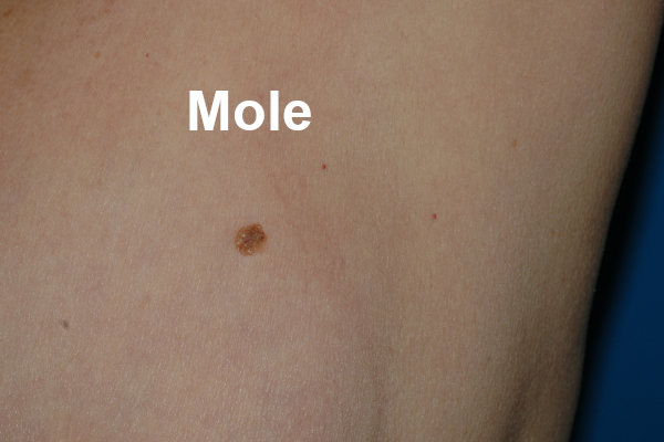
Particularly, moles on the face can cause discomfort in terms of number and appearance, and their treatment is surgical. In addition, benign tumors can cause physical problems in places such as eyelids, lips, nose, genital area, and surgical treatment is applied for functional purposes.
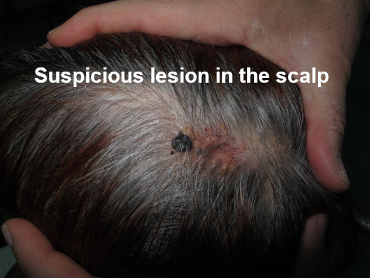
Surgery for potentially malignant lesions
Various findings suggest that a skin lesion may be malignant. These:
- Sudden growth of an existing lesion and starting to appear new similar lesions in its neighborhood.
- Change of color.
- Change in shape and texture
- Bleeding
- Appearance of sudden growing new lesion.
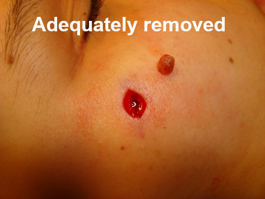
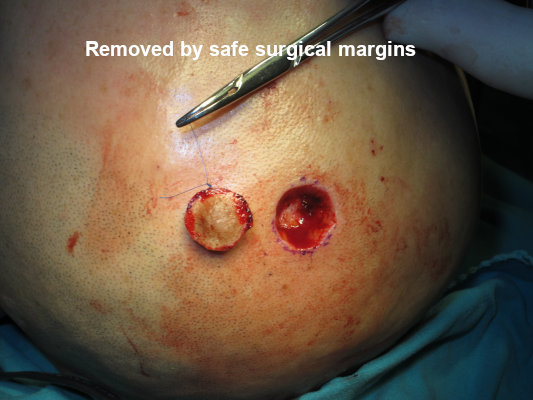
The most guaranteed treatment for any suspicious skin lesion is to remove it and send it to pathology. The absolute requirement to safely remove a lesion is to remove the entire lesion, both in the edges and depth. It might be visible to the eye how far the edges of a lesion extend. However, in reality, the lesion may have progressed beyond the visible parts in depth. Therefore, each lesion must be removed from a distance called the safe surgical margin.
What are safe surgical margins?
Benign skin tumors often extend no more than 1 mm beyond their visible margins. For this reason, it is sufficient to remove a nevus (mole) that we think is benign, including 1 mm of intact skin from its visible edges. However, in malignant skin tumors, the surgical margin, that is, the intact skin and subcutaneous tissue that needs to be removed, varies according to the type of tumor. This margin may be 0.5 cm in moderately malignant tumors, while this border may be up to 5 cm in advanced malignant tumors. Plastic Surgeons are faced with a problem in tumors whose diagnosis is doubtful. There is a very important difference between removing a tumor with 2 mm of intact skin and removing it with 5 cm of intact skin. If a tumor of unknown origin is 1 cm or less in diameter, all of it is removed with a surgical margin of at least 2 mm and sent to pathology. If the lesion is benign in the report, there is no need for additional treatment. If the lesion is malignant in the report, the following should be noted:
- If the surgical margins are clean, it means that there is no visible tumor left behind. The type of tumor is considered, and the treatment is considered sufficient in moderately malignant tumors and the operation area is monitored intermittently for any recurrence. If there is a recurrence, surgery is performed again. If the tumor report is an advanced malignant tumor, although it seems that nothing is left behind, the surgical margin of 2 mm is insufficient and wider surgery and additional treatments are performed on the same area without waiting.
- If there is a tumor within the surgical margins, the surgery was definitely inadequate and a more appropriate surgical treatment is performed immediately.
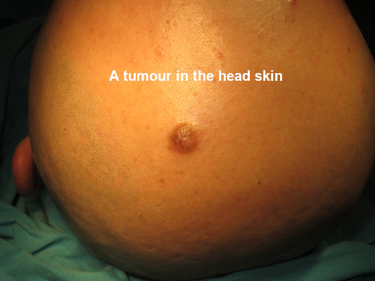
Surgical treatment of suspicious skin lesions with a large diameter
Suspected lesion is a lesion that is unknown whether it is benign or malignant. If it is known to be benign, surgery is not necessary. On the other hand, if it is malignant, it will be removed with some healthy skin around it. Since the already large lesion will be removed much larger, the diameter of the wound (defect) will also be large and additional procedures will be required to close it. In this case, biopsy(s) are taken from the suspicious lesion. A biopsy means taking a small piece(s) from a lesion so that no additional action is required to close it. Most of the time, these specimens are taken under local anesthesia in outpatient offices and immediately sent to pathology. Appropriate treatment is planned according to the forthcoming pathology report and then performed.
Closing the wound that occurs after removing a skin lesion
When a part of the skin is cut and removed, the resulting gap is closed with different methods.
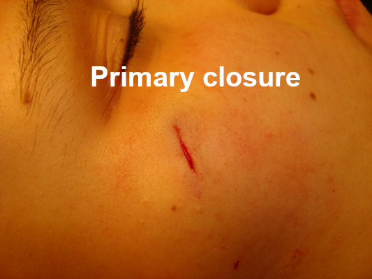
Primary closure
The skin has elasticity. For this reason, skin deficiencies up to 1 cm in diameter in many areas are pulled to the middle, and the opening is removed. Skin defects with a diameter of 3 cm or larger, depending on the area of the skin, can be closed primarily, that is, by suturing side by side.

Closure with skin graft
If the defect that occurs after the removed tumor is wide, then the skin edges cannot be joined in the middle. In this case, the opening is closed with a skin patch (graft) taken from another area.


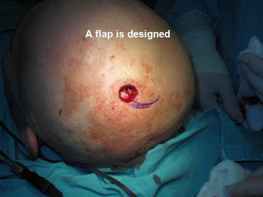
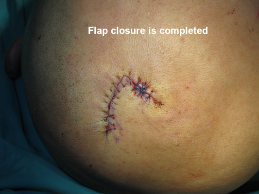
Closure with skin flaps
If there are important structures (vessels, nerves, bones, cartilage) on the exposed wound surface after the skin lesion is removed, they should be covered with a solid and thick layer of tissues. In this case, skin flaps are used.
Conclusion
Skin lesions can be removed for aesthetic or health reasons. Each lesion should be sent to pathology after removal and it should be learned what the lesion is and whether the surgical margins are clean. Treatment of skin tumors is the job of Plastic Reconstructive and Aesthetic Surgery.

